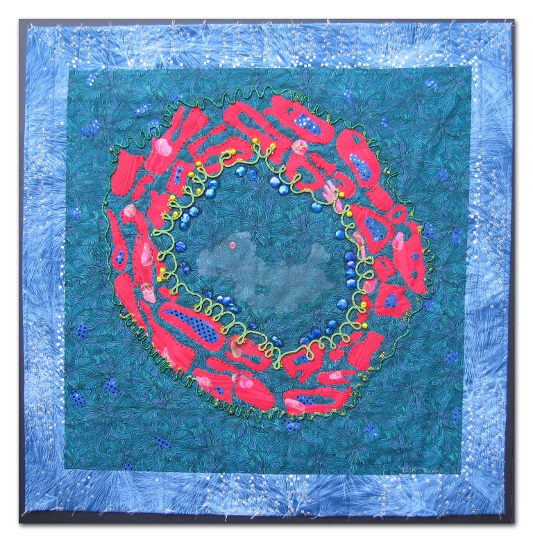
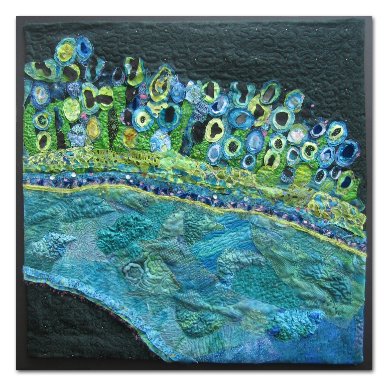
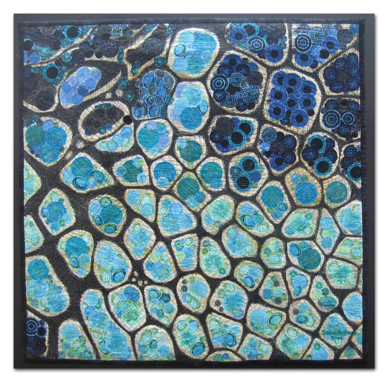
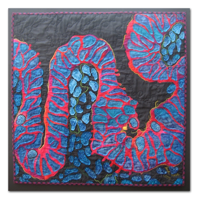
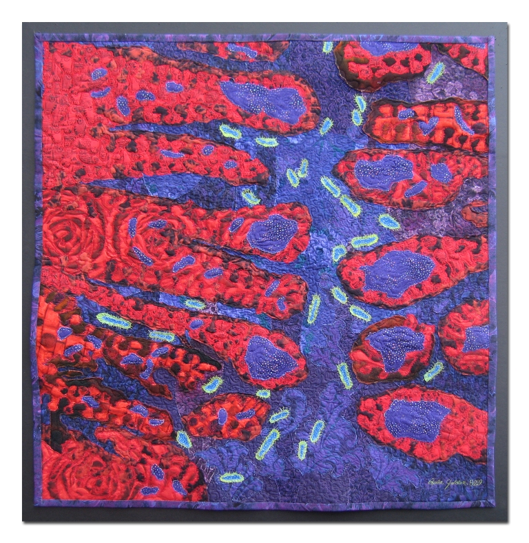
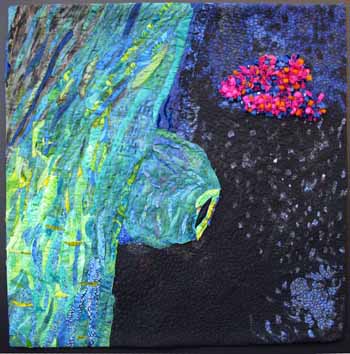
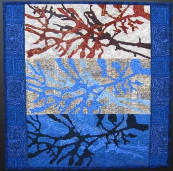
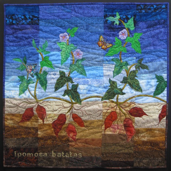
 Arterial Dream by Judy Busby Arterial Dream by Judy Busby
This quilt is a rendition of Greg Dressler’s photograph of a section through a major blood vessel. The background is machine quilted with hand sewn embellishments that include: Czech glass beads, Swarovski crystals, marbled beads and embroidered French knots. Layers of tulle and satin fabrics are hand sewn with raw edge appliqué. Leather and woven cords are hand couched. As Greg Dressler and his fellow researchers pass by this quilt, I hope they will feel the respect and thankfulness I feel for their dedication and talent! |  Gregory Dressler, Ph.D., Professor, Pathology, Gregory Dressler, Ph.D., Professor, Pathology,
Co-Director, Center for Organogenesis, University of Michigan
This is a section through a major blood vessel stained to observe the smooth muscle (red) and a structural protein in the supporting basement membrane (green). The cell nuclei are stained blue. |
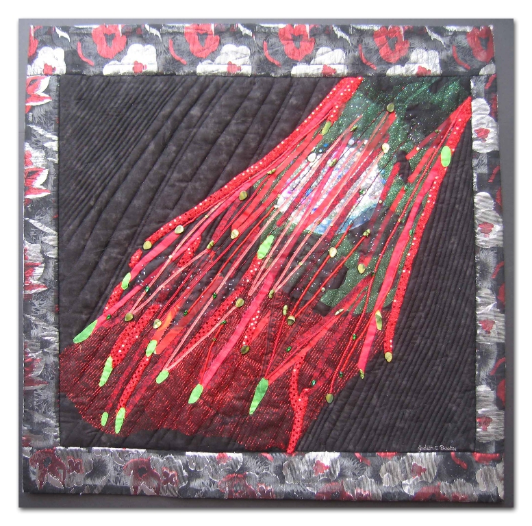 Fibroblast by Judy Busby Fibroblast by Judy Busby
My quilt is a rendition of Michael Dame’s photograph of a fi broblast – a human skin cell labeled with various dyes distinguishing the proteins that allow the cell to adhere and to spread. The background is machine quilted with hand sewn embellishments that include: Czech glass beads, fiber optic beads, tear drop shells, and Betel nut halves. Layers of tulle, satin ribbons, and various fabrics are hand sewn with raw edge and turned under appliqué techniques. As Michael Dame and his fellow researchers pass by this quilt, I hope they will feel the respect and thankfulness I feel for their dedication and talent! |  Michael Dame, Research Associate, Michael Dame, Research Associate,
Department of Pathology, University of Michigan
This human skin cell, a fibroblast, is growing in a culture and has been labeled with dyes that identify the proteins that enable the cell to attach and spread (green – vinculin; red – actin). Skin fibroblasts secrete proteins that anchor the cells and allow them to spread. Studying the way cells attach and spread helps us understand how cells behave in skin diseases. |
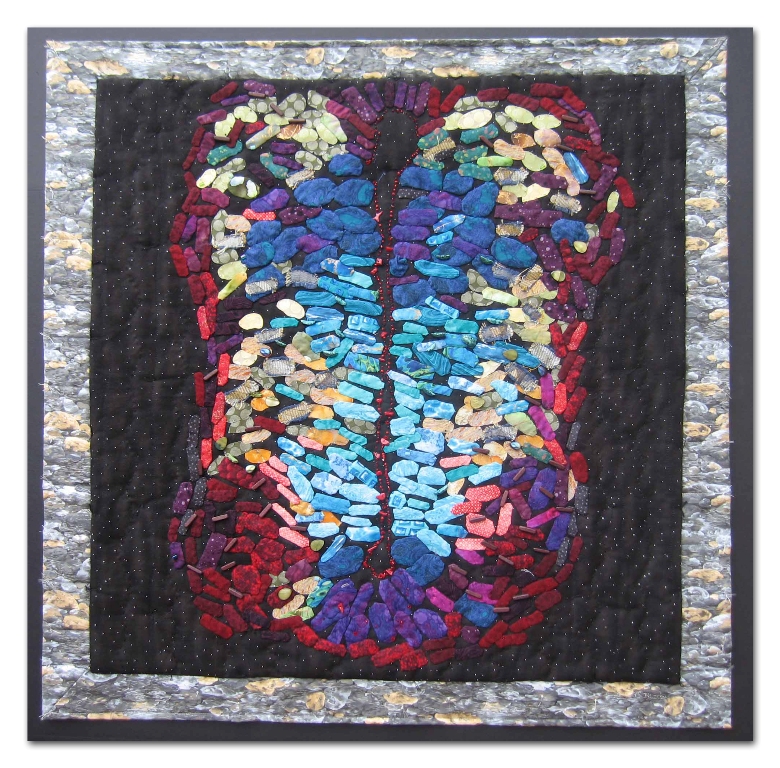 Rainbow Trout by Judy Busby Rainbow Trout by Judy Busby
This quilt is a rendition of Zachary Gaber’s photograph of a cross section of a four-day-old chick embryo’s spinal cord. The background is hand quilted and machine quilted. Hand sewn embellishments include: Swarovski crystals, Czech glass beads, fiber optic beads, red coral, Betel nut halves and wooden beads. Both raw edge and turned under appliqué techniques are used to create the individual cells. As Zachary Gaber and his fellow researchers pass by this quilt, I hope they will feel the respect and thankfulness I feel for their dedication and talent! |  Zachary Gaber, Graduate Student, Zachary Gaber, Graduate Student,
Cell & Developmental Biology, University of Michigan
The developing spinal cord contains populations of neural progenitor cells that
form the nerves and supporting cells of the adult spinal cord. This image shows
a cross-section of a four-day-old chicken embryo at the time when the progenitor
cells are just beginning to form nerve cells. The cyan, blue, and turquoise cells
at the center of the spinal cord are different classes of progenitors. The cells
maturing into nerves are stained yellow, orange and red. Although this is a
picture of a chick embryo, a mammalian embryo would look quite similar. |
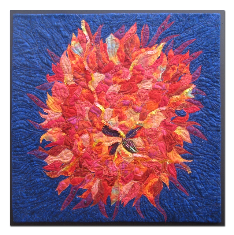
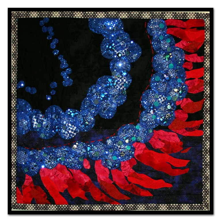 Fire in Her Eyes by Judy Busby Fire in Her Eyes by Judy Busby
This quilt is a rendition of Rebecca Bernados’s photograph of a section of a zebrafish retina. The blue spots are created with sequins covered with fabric and sewn by hand. The varying levels of circles are individually hand sewn, giving depth to the band of nuclei. The red photoreceptors include red fabrics and handmade paper. The middle, red portion is depicted using shades of red glass beads. As Rebecca Bernados and her fellow researchers pass by this quilt, I hope they will feel the respect and thankfulness I feel for their dedication and talent! |  Rebecca Bernardos, Graduate Student, Rebecca Bernardos, Graduate Student,
Neuroscience Program, University of Michigan
Degeneration of cells in the retina is the most common cause of irreversible blindness in the Western world. When the cells that detect light (photoreceptors) die in the human retina, they are never replaced. Some fish, on the other hand, have the remarkable ability to produce new photoreceptors after injury. The goal of our research is to understand how these cells regenerate in the fish retina with the hope that this will suggest ways to reverse the effects of retinal degeneration in humans. This photomicrograph features a section of a zebrafish retina. The red feather-like cells are photoreceptors and each blue spot is a cell nucleus. |
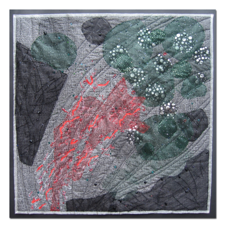
 Edge of the World by Annabel Ebersole Edge of the World by Annabel Ebersole
This stunning photograph grabbed my attention right away. Our stomach is integral to good health! Here was an ethereal picture that I could interpret in fabric and thread. Creating the textures with a new product “Texture Magic” made the bumpy looking parts of the bottom section come out just right. For the top half, I cut out many oblongs of different textures and stitched them together before they were applied to the backing. Using silk, wool, cotton, velvet and organza made these cellular structures look like the photo. Finally, quilting, painting and beads gave me the iridescence I hoped for. |  Deb Gumucio, Ph.D., Director, Center for Organogenesis, Deb Gumucio, Ph.D., Director, Center for Organogenesis,
Professor, Cell & Developmental Biology, University of Michigan
This image shows the posterior part of the stomach, a region called the antrum. The antrum is important because the cells that reside here control the release of acid from the more anterior stomach. They also direct the release of enzymes from the pancreas that are needed for absorption as food moves into the intestine. This control point is also one of two regions of the stomach that are highly susceptible to cancer. Stem cells that reside deep within the flask-like green structures are responsible for the regular renewal of this surface. Those same stem cells are also believed to be the most likely source of stomach cancers. |
 Castor & Pollux by Annabel Ebersole Castor & Pollux by Annabel Ebersole
Fireworks in a mouse stem cell! Who could imagine this? What fun I had in painting and stamping the surface, adding layers of fuchsia, cobalt and red violet. Next more layers, hand stitching, machine quilting and the final wispy lime and fuchsia “zingers!” Now the structures dance! |  Nicole Slawny, Graduate Student, Nicole Slawny, Graduate Student,
Cell & Developmental Biology, University of Michigan
Mouse embryonic stem cells were engineered to overexpress a protein called geminin. This protein has two important functions during embryonic development: to control cell division and to promote differentiation of neural cells. The geminin-producing cells form many immature neural precursors (purple) and neurons (green). The cell nuclei have been stained blue. Experiments with embryonic stems cells are important not only to produce specific cell types for cell replacement therapies, but also as a model system to understand embryonic development. |
 Green Feather by Annabel Ebersole Green Feather by Annabel Ebersole
This quilt is based on the photo of a lobule of the cerebellum, enhanced with staining. The green astrocytes support the large specialized neurons or red Purkinje cells. Neural stem cells are some of the cells that are stained blue. The quilt uses fabric and fiber to represent parts of the cells of the cerebellum. Silk throwster’s waste, silk cocoon fibers, silk Sari yarn from Nepal, glitter threads, cotton, rayon and silk threads, stamp pads, paint, stencils, wool yarn, hemp string, rubber string, metallic grid and various fabrics, both hand and machine stitched, were used to create these effects. |  Maria Morrell, Ph.D., Postdoctoral Research Fellow, Maria Morrell, Ph.D., Postdoctoral Research Fellow,
Cell & Developmental Biology, University of Michigan
Located at the lower back of the brain, the cerebellum is a fist-sized structure that contains more neurons than all of the rest of the brain combined. The cerebellum performs a variety of roles, controlling motor, sensory, cognitive, and linguistic functions. In this section of a highly folded lobule, astrocytes (green) support large specialized neurons, the Purkinje cells (red). The blue nuclear staining illustrates the many cells present in each of the lobules. Cerebellar neural stem cells are located next to the Purkinje cells. |
 Escher’s Needlepoint by Lisa Ellis Escher’s Needlepoint by Lisa Ellis
I was immediately drawn to this photo by Kaelyn Male when browsing the collection. I loved the colors and the composition. Who knew that the gut could be so beautiful! As a Mathematician, I am always drawn to Escher’s works and could see why this piece was so named. I enjoyed playing with embellishments to try to capture the sparkle of the green and the three dimensional look of the purple. I used paint, Angelina fibers and metallic bits for the inside of the cells. |  Kaelyn Male, Graduate Student, Cell Biology, Duke University Kaelyn Male, Graduate Student, Cell Biology, Duke University
The surface of the gut is thrown into small projections (villi) with invaginations (crypts) at their base. The villi help to increase the area of the gut surface for absorption, while the crypts are the home of the stem cells that are responsible for continuous renewal of this epithelium. This is a cross-section of villi and crypts in the small intestine of an adult mouse. It is stained to identify cell nuclei (purple), and to show junctions between cells (green). |
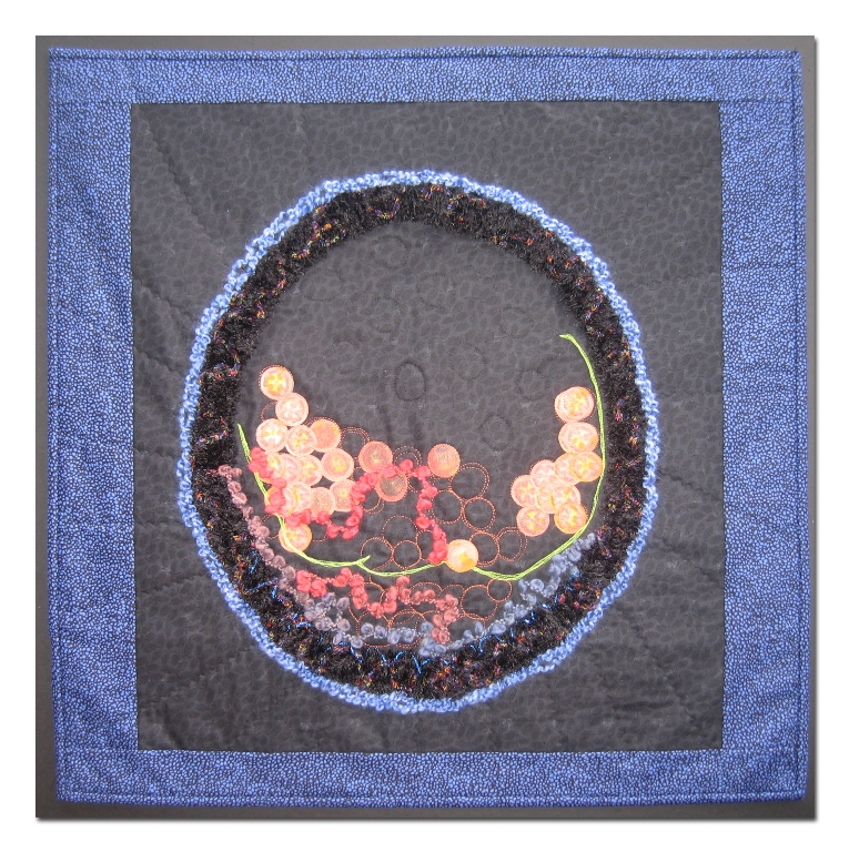
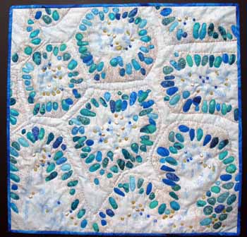
 That’s Phernominal by Sandi Goldman That’s Phernominal by Sandi Goldman
My inspiration was Louise Hecker’s BioArtography piece That’s Phernominal. I used part of the original image and painted the black line work on a piece of batik fabric. The next step was to add the circles, either cut from commercial fabrics or drawn on, and then the whole piece was machine quilted and more circles were drawn on. I was initially attracted to this image because of the coloration. I enjoy this color palette, and find I use it in many of my quilts. I am currently working on a series of quilts using circles; I like their rhythm, balance and continuity. |  Louise Hecker, Graduate Student, Louise Hecker, Graduate Student,
Cell & Developmental Biology, University of Michigan
One of the amazing ways in which animals communicate is by pheromones. In the red-backed salamander (Plethodon cinereus), these chemicals are released from skin glands in the tail and play a role in species recognition, territory marking and mating behavior. In this photomicrograph, cells synthesize pheromones (turquoise) and pack the aromatic chemicals into small oil droplets. By studying pheromonal communication in other animal species, we can gain a better understanding of how humans might unwittingly use this evolutionarily ancient form of communication. |
 Visibly Complex by Christine Adams Visibly Complex by Christine Adams
My love of fabric and its varied possibilities began as a little girl in my grandfather’s upholstery shop. My intent with Visibly Complex was not to render a realistic scientific representation, but to convey the flavor of the image. This project was one of the few times I’ve worked with whole piece quilting – allowing free motion quilting to tell the story. I dyed and painted the fabric both before and after stitching. I also used poetic license in coloring – bleeding browns to pink and gold and sometimes green. This was an exciting process. I feel the stitching strengthens the piece – the dye and paint are an embellishment. |  Tom Glaser, Associate Professor, Tom Glaser, Associate Professor,
Department of Internal Medicine, University of Michigan
This retina of a laboratory mouse is viewed with Nomarski optics and stained with an antibody to identify ganglion cells, cone photoreceptors and the inner plexiform layer (brown). A quarter of a millimeter across, this layered tissue covers the inside surface of the eye. The human retina is similar, and functions as a highly organized switchboard that records, processes and conveys all visual information from the outside world to the brain. In this image, light first passes through the lens and then enters the retina to excite the cone and finger-like rod photoreceptors located at the back of the eye near the pigmented epithelial layer (black). The resulting electrical signals are then relayed stepwise, via delicate neural tendrils, to the ganglion cells, whose projections join to form the optic nerve and travel to the brain. |
 Inner Beauty by Barbara Hollinger Inner Beauty by Barbara Hollinger
A bundle of glowing energy, this sea urchin embryo was added layer by layer onto a single piece of cloth. The web of color was created using a resist of school glue with each darkening shade of color applied, one after the other. Once the glue was washed away, the structure of the embryo remained. More layers of thread, yarn, beads and paint bring to life the creature as it floats across the textured background. |  Kate Walton, Graduate Student, Cell Biology, Duke University Kate Walton, Graduate Student, Cell Biology, Duke University
The inner beauty of the sea urchin embryo is revealed here in multiple colors. The skeleton (shown in turquoise) is composed of individual cells that become mineralized to provide the outer framework of the embryo. Supported by the skeleton, the gut of the embryo is shown in yellow and the muscle is stained purple. The elaborate organization of the muscle can be seen in the perfectly organized striations of muscle surrounding the foregut, which allows the embryo to swallow food. The outline of the gut sphincters can be seen in pink. At the tip of the embryo, there are 21 muscles that do not have a known function but are suspected to help the embryo steer as it swims in search of food. |
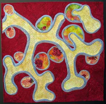
 Guts & Glory by Carole Nicholas Guts & Glory by Carole Nicholas
The colors of Guts and Glory are the reason I chose to interpret this image in fabric. I like how the apple greens shade to sunlit yellows and gold, then blend into dazzling oranges. They seem to glow on the dark background. I thought it would be a challenge to try to mimic the effect of the cells floating by in a brilliant citrus stream. |  Lymari Lopez-Diaz, Ph.D., Postdoctoral Research Fellow, Lymari Lopez-Diaz, Ph.D., Postdoctoral Research Fellow,
Department of Molecular and Integrative Physiology, University of Michigan
The job of the intestine is to absorb food. To provide maximum surface area, the absorptive surface of the intestine is shaped into billions of finger-like projections called villi. This is a cross-section of one villus. The absorptive epithelial cells are on the outside of the villus (green). The orange and yellow cells in the center include blood vessels, nerves, muscle and immune cells, all of which function with the epithelial cells as a cohesive unit designed for absorption. |
 Loch Ness by Carole Nicholas Loch Ness by Carole Nicholas
The photograph of actively dividing intestinal cells, despite its title, does not conjure up an image of a deep, cold loch with a mysterious, mythical monster lurking in its murky depths. Rather, it looks to me like a joyous celebration, with a colorful storybook character Nessie emerging to engage in some magic or whimsy. I used a Scottish highlands tartan for the binding of the quilt. The background is moiré taffeta, to add texture and movement. The cells are mostly dyed silk chiffon and some cotton. Glass beads and metallic and rayon threads were used as embellishment. |  Åsa Kolterud, Postdoctoral Research Fellow, Åsa Kolterud, Postdoctoral Research Fellow,
Cell & Developmental Biology, University of Michigan
During embryogenesis, the developing intestine undergoes a remarkable remodeling
process in which the surface of the gut tube is folded into finger-like projections
called villi. These villi, which extend into the lumen of the gut tube, drastically
increase the absorptive surface of the intestine and are important for efficient
nutrient uptake. This photomicrograph shows the initial buckling of the intestinal
epithelium (stained red) into nascent villi. The nuclei of the cells are stained blue.
Note the flower-like nuclei within the epithelium – these are dividing cells lining
up their chromosomes. |
 Crystal Ball by Donna DeSoto Crystal Ball by Donna DeSoto
Who knew that an urchin embryo could produce something as dynamic as the photograph that was the basis for my artwork? I love the colors and also the shapes and 3-D effect. I used a number of cotton and synthetic fabrics and fibers, raw-edge appliquéd, free-motion stitched and hand embellished. Photo transfer was used to depict the cells that will form connective tissue. This quilt is dedicated to the memory of my friend, Patricia Botta. |  Esther Miranda, Research Analyst, Cell Biology, Duke University Esther Miranda, Research Analyst, Cell Biology, Duke University
Blue stains the outside of the urchin embryo almost like a cage in a 3-D projection, while the cells inside that will eventually form the skeleton are stained red. The green connecting fibers hold the cells together like clusters of grapes, while the bright yellow cells will form connective tissue. This complex structure provides a beautiful crystal ball for developmental studies. |
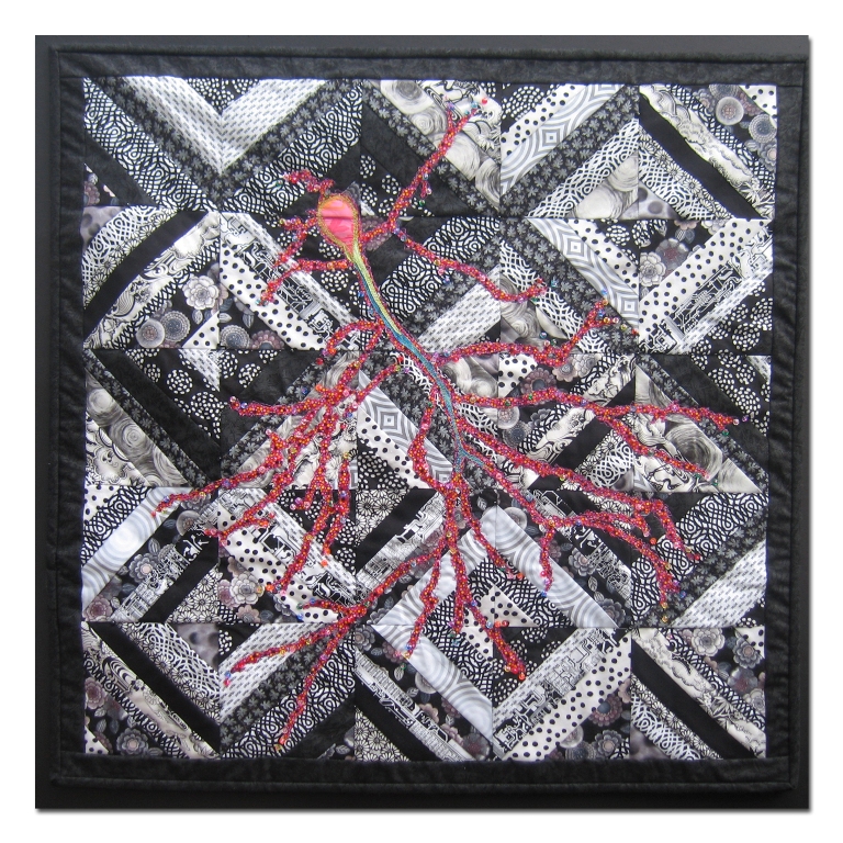 Eureka by Donna DeSoto Eureka by Donna DeSoto
I have to admit that the word “hippocampus” makes me smile! When I first heard of the website containing BioArtography images, I studied each one and read each description. As soon as I found out that the subject of one of the images includes the area of the brain that is involved in memory, I knew that I wanted to make that quilt. The idea for the black and white background of this piece came from Barbara Persing’s book called StrataVarious Quilts. Materials used were commercial cottons; the raw edge design was free motion quilted and glass beads were applied by hand. |  Benjamin Carlson, Graduate Student, Cell Biology, Duke University Benjamin Carlson, Graduate Student, Cell Biology, Duke University
This image is of a mouse neuron from the hippocampus, the brain area that is critically involved in memory. The large branching projections contain spiky spines at their tips that receive signals from other neurons. These spiny structures are thought to be involved in the processing of information during learning and memory function. In this image, the neuron is stained to identify a protein that forms the internal skeleton of the cell. This allows us to study the structure of these spines during memory formation and learning. |
 Gutsy by Donna DeSoto Gutsy by Donna DeSoto
Two things initially drew me to this photo: the beautiful blue colors and the organic shapes! I have an extensive collection of all shades of blue fabric, and I was eager to use it, set off against a stark black background. This piece was constructed with commercial cotton fabric, some enhanced by the addition of paint and/or stamping. Raw edged shapes were applied to the background by free motion quilting. This quilt was made in honor of my father-in-law, Oscar DeSoto, who was born in Cuba. |  Blair Madison, Ph.D., Postdoctoral Fellow, and Andrea Waite, Blair Madison, Ph.D., Postdoctoral Fellow, and Andrea Waite,
Graduate Student, Cell & Developmental Biology, University of Michigan
The surface of the intestine is lined by millions of finger-like structures (villi) that extend into the lumen of the intestine to provide enormous surface area for absorption of nutrients. This photomicrograph shows a portion of an intestinal villus. The blue ovals are nuclei of individual cells that cover the villus’ surface. These cells absorb proteins, sugars and fats from food in the gut lumen. The absorbed nutrients are processed by these cells, and then secreted into blood vessels that spiral up into the center of the villi (pink). The muscles (beige) in the center squeeze the villi to pump nutrients into the main blood stream. |
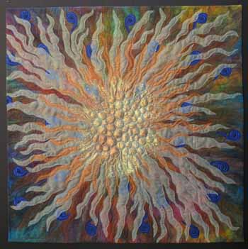
 Sunburst by Bunnie Jordan Sunburst by Bunnie Jordan
This is my interpretation of a micrograph of neural stem cells. A prism design and a class from the artist Barbara Olson was my starting point. The techniques I used include fused and machine appliqué incorporating yarns, organza, netting and beads. |  Maria Morrell, Ph.D., Postdoctoral Research Fellow, Maria Morrell, Ph.D., Postdoctoral Research Fellow,
Cell & Developmental Biology, University of Michigan
The goal of our research is to study the potential of neural stem cells for repair after injury to the central nervous system. Certain regions of the adult brain contain neural stem cells that have the capability to form all types of neural cells: neurons (nerve cells), astrocytes (supporting cells) and oligodendrocytes (myelin forming cells). In this photomicrograph, the spherical cluster of neural stem cells was stained to identify a protein typically found in astrocytes (red). The nuclei of the cells have been stained blue. |
 Contact by Judy Busby Contact by Judy Busby
My quilt is a rendition of Catherine Krull’s photograph showing the reaching of “long processes” of neurons from a developing spinal cord towards lower limb muscles. The background is machine quilted with hand sewn embellishments that include: Czech glass beads, Swarovski crystals, marbled beads and embroidered French knots. Cotton, satin and tulle fabrics are hand sewn with raw edge appliqué. Acrylic paint with textile medium is used as highlighting. Sashiko and hand stitching are included. |  Catherine Krull, Assistant Professor, Catherine Krull, Assistant Professor,
Cell & Developmental Biology, University of Michigan
Neurons send their long processes from the developing spinal cord to contact muscles in the lower limbs. As these processes extend towards their muscle targets, they encounter proteins in the environment that influence their growth. Here, the growing processes (red) stop at the base of the lower limb, where they reach ephrin proteins (green). |
 Goblins by Paula Golden Goblins by Paula Golden
Through the use of texture, I explore the duality of nature in the work I create. Within the realm of each dimension, each form is meaningless without an appreciation of its relationship to its complement, whether it be of ideas, colors or images.
The connectedness to times past and present is an important variable in my choice and use of materials. I call this piece IBD. I-B-D, three simple letters, but the impact the disease they represent (Inflammatory Bowel Disease) greatly alters a person’s life. New medications, alternative mind-body approaches and research offer promise for the future. |  Åsa Kolterud, Ph.D., Postdoctoral Fellow, Karolinska Institute, Sweden Åsa Kolterud, Ph.D., Postdoctoral Fellow, Karolinska Institute, Sweden
The colon is made of billions of flask-like structures called glands (shown in red). In colon cancer, tumors develop from these epithelial cells. The green/aqua cells in the center of this picture are part of the connective tissue. Many of these cells are immune cells that function to protect the colon from invasive bacteria. The immune cells and epithelial cells communicate with each other using soluble proteins as messages. We are trying to understand the nature of this cellular
cross-talk. In inflammatory bowel disease (Crohn’s disease or ulcerative colitis), this communication is compromised and proteins secreted by the normally protective immune cells damage the epithelial cells. |
 Learning Tree by Christine Adams Learning Tree by Christine Adams
I used to think that life was like fabric that can be arranged and rearranged so that each time the same yardage tells a slightly different story. My grandfather allowed me freedom to explore his upholstery shop and its scrap box. Working on the Learning Tree was an extension of these happy days. To bring out the richness and depth of the background, I chose suede upholstery fabric dyed charcoal and then overdyed with various shades of blue. Silk waste dyed red, net, and sequins were used to complete the image. Unusual tools used in this project were an ice pick and a leather punch. |  Maria Morrell, Ph.D., Postdoctoral Research Fellow, Maria Morrell, Ph.D., Postdoctoral Research Fellow,
Cell & Developmental Biology, University of Michigan
This is a section of a mouse cerebellum (from the Latin “little brain”). Located
at the lower back of the brain, the cerebellum receives and sends information to other organs. Accordingly, this information processing center contains more than 50 percent of the total neurons in the brain. To accommodate such a large number of cells into such a small space, the outer region of the cerebellum is thrown into folds, or lobules. In this photomicrograph, every red dot indicates one single cell that is undergoing cell division. Conditions such as schizophrenia, autism, mood disorders, dementia, and attention deficit hyperactivity disorder (ADHD) are all thought to be disorders of cerebellar function. |
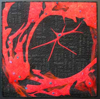
 Calimari by Annabel Ebersole Calimari by Annabel Ebersole
Nerve studies resonate with me as my Dad died from Alzheimer’s. Our human body is an amazing working universe and these studies that illuminate possible “fixes” are filled with hope. I have painted the background with colored pencils, oil paint sticks and liquid paint. Using wool, silk, cotton, ultra suede and rayon fabrics has made the long blue green fibers and pinky- explosive spot stand out. Bravo to these research efforts!! |  Calimari by Andy Chervenak, Graduate Student, Cell and Developmental Biology Calimari by Andy Chervenak, Graduate Student, Cell and Developmental Biology
The green structure in this picture is the siphon of a marine squid. The siphon sucks water into the squid and then rapidly expels it to propel the squid through the ocean. Surrounding muscles can change the direction of the jet of water, so that the squid can control its direction. Thus, the squid is a wonderful model for the study of muscle development and function, but it is more well known for another specialization, its giant axon, or nerve fiber, that is 1,000 times wider than the axons present in humans. For nearly a century, scientists have studied the squid giant axon to understand how nerves conduct electricity and how nerve cells communicate with muscle and with other nerve cells. Disturbances in these nerve communication functions are responsible for the symptoms in Alzheimer’s Disease and other adult-onset neurodegenerative diseases, such as Parkinson’s and ALS. |
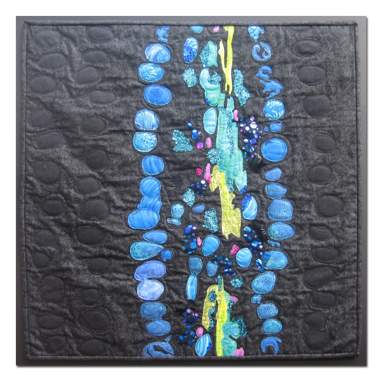
 Aztec Villi by Donna DeSoto Aztec Villi by Donna DeSoto
We’ve just experienced one of the hottest summers on record in the DC metropolitan area. No wonder that this image drew me in: working with various hand-dyed and cotton fabrics of many hues of blue-green and turquoise in my air conditioned home was a welcome respite from the heat and humidity outside. I used Caran d’Ache Neocolor Aquarelle Crayons to achieve effects of the blurred epithelial cells, and highlighted areas of villi with Jacquard Lumiere fabric paint and glitter. This piece is dedicated to our good family friend, John Dreska, and my quilting buddies Mary Lois Davis, Betsy Smith and Dana Hancock, who all suffer from Celiac disease. |  Aztec Villi by Aaron Udager, MD/PhD Graduate Student, Cell and Developmental Biology Aztec Villi by Aaron Udager, MD/PhD Graduate Student, Cell and Developmental Biology
This is an image of a fetal mouse intestine. The brown cells with blue nuclei are intestinal epithelial cells. In the adult animal, these cells will be responsible for the processing and uptake of nutrients in the gut. Because the intestine needs a huge surface area for proper absorption, the epithelium is thrown into folds, called villi. Stem cells are located at the base of each villus and are responsible for replacing the entire epithelium every 3-4 days. The final surface area of the intestine in an adult human is approximately 2,000 square feet, bigger than many houses! Loss of villi occurs in Celiac disease, leading to malabsorption. |
 Thistle by Barbara Hollinger Thistle by Barbara Hollinger
The hand dyed and hand painted background has been overlaid with several iridescent layers of sheer fabric, threads and fibers allowing the flash of color and light to come through hoping to capture the electricity pulsing between these neural cells sending each other signals for motion, for thought, of joy and of pain. |  Thistle by Maria Morrell, Ph.D., Postdoctoral Research Fellow, Cell and Developmental Biology Thistle by Maria Morrell, Ph.D., Postdoctoral Research Fellow, Cell and Developmental Biology
The goal of our research is study the potential of neural stem cells for repair after injury to the central nervous system. Certain regions of the adult brain contain neural stem cells that have the capability to form all types of neural cells: neurons (nerve cells), astrocytes (supporting cells) and oligodendrocytes (myelin forming cells). In this micrograph a spherical cluster of neural stem cells was stained to identify protein typically found in neurons (green) or astrocytes (orange). The nuclei of the cells have been stained blue. |
 Curious by Mary Lois Davis Curious by Mary Lois Davis
My inspiration was Maria Morrell’s photograph of “Curious” with its fabulous red/orange on black coloring with little specks of blue, green and pink. The black background of this piece has screen-printed words taken from Dr. Morrell’s description of the slide: brain, glia, cell nuclei, neural stem cells, and so on. The red cell structures were created with raw-edge appliqué and free motion stitching along with needle felting to apply wool roving to create the wispy bits. Some of the red stitching was done using hologram thread. The colorful specks are represented with bead clusters. |  Curious by Maria Morrell, Ph.D., Postdoctoral Research Fellow, Cell and Developmental Biology Curious by Maria Morrell, Ph.D., Postdoctoral Research Fellow, Cell and Developmental Biology
Adult neural stem cells are present in several discrete locations in the brain. We are interested in how these cells can be directed to replace neurons and glia (supporting cells) lost due to injury, disease or normal aging. These neural stem cells were removed from an adult mouse brain, grown in tissue culture and induced to differentiate into glial cells; the supporting cells of the nervous system. The processes of the many glial cells are red. The round blue structures are cell nuclei. |
 Van Gogh’s Skin by Carole Nicholas Van Gogh’s Skin by Carole Nicholas
“Van Goghs Skin: The photograph under the microscope which inspired this piece surely is reminiscent of the now iconic Van Gogh’s”The Starry Night”. I tried to imitate in fabric and stitching Van Gogh’s bursts of brushwork to create all the swirling movement he captured with paint. |  VanGogh’s Skin by Mark Hutchin, House Officer, Dermatology VanGogh’s Skin by Mark Hutchin, House Officer, Dermatology
This image shows a basal cell carcinoma, the most common form of skin cancer. The top portion shows the normal skin surface of a mouse, which stains red with this special stain (Masson trichrome stain). The blue is collagen, a strong fibrous substance that provides structural support for the skin. The appearance of human skin is very similar to this. At the bottom of the image, red staining tumor cells are seen. The yellow has been added for visual interest by the artist. By studying the development of tumors in the mouse, we hope to learn how to prevent the formation of similar tumors in humans. |
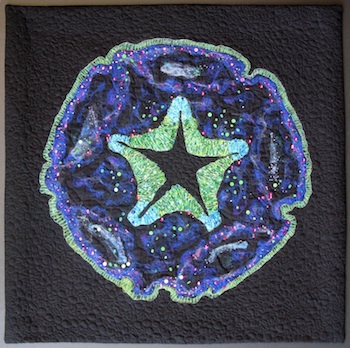 Stem Star by Lisa Ellis Stem Star by Lisa Ellis
This image spoke to me with its bright colors and similarities with creatures one might find down at the beach at our summer home in California. I used a combination of needle punch felting, bobbin thread painting and a variety of threads and beads. |  Stem Star by Shannon Davis, Ph.D., Research Investigator, Human Genetics Stem Star by Shannon Davis, Ph.D., Research Investigator, Human Genetics
This is a section of a mouse pituitary gland and nearby neural tube that has been artificially transformed into a star pattern. The neural tube forms the central star, while the pituitary is the bottle-cap shaped structure on the outside of the “stem star”. The name, Stem Star, was given because both the neural tube and the pituitary contain stem cells that are responsible for the growth and maintenance of the two organs. The pituitary gland actually derives (in part) from the neural tube; the pituitary is a master gland that controls the activity of other glands that regulate growth, pregnancy, water balance, energy metabolism, blood pressure and the body’s response to stress. |
 Mellow by Sandi Goldman Mellow by Sandi Goldman
I could instantly see the creative possibilities in “Mellow”. I was connected to the circles, the texture, the rhythm, and the soft colors in the image. But when do I stop the embroidery, the painting, the drawing-is it finished? When does the research end? When will there be a cure, a breakthrough, an answer? This exemplifies the link between art and life, the intertwining of research and art. |  Mellow by Matthew Velkey, Ph.D., Lecturer, Cell and Developmental Biology Mellow by Matthew Velkey, Ph.D., Lecturer, Cell and Developmental Biology
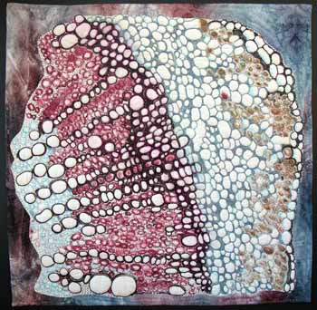
Some of the first studies of cells were done in plant tissues. In fact, Robert Hooke first coined the term “cell” in 1665 based on his examination of thin slices of cork under the microscope. Hooke observed thousands of tiny compartments within the cork tissue reminiscent of the tiny rooms, or cells, of a monastery. This image shows a stem of the industrial hemp plant, Cannabis sativa, cut in cross section, revealing the numerous vessels, made up of cells connected end-to-end, that transport nutrients and water. The section is NOT stained, so the colors present are due to natural pigments in the plant. The stem of a hemp plant contains an inner, woody pith, or hurd, made up of xylem vessels (red), which transport water. The outer layer, or bast, contains phloem vessels (aqua) that transport sugars made in the leaves. The bast fiber bundles (greenish-red bundles) located in the outer layer are very strong and are used to make hemp products such as textiles and rope. This subspecies of Cannabis has a relatively low concentration of tetrahydrocannabinol, the psychoactive component found in marijuana. |
 Internet by Judy Busby Internet by Judy Busby
My quilt is a rendition of Maria Morrell’s research photograph of adult neural cells of mice. I am thankful to have permission to use Ms. Morrell’s remarkable photograph! The background is machine quilted with hand-sewn embellishments that include: Czech glass beads, Swarovski crystals, marbled beads and embroidered French knots. Layers of tulle and satin fabrics are hand sewn with raw edge appliqué. As Ms. Morrell and her fellow scientists view this artwork, I hope they will feel the honor, respect, and thankfulness I feel for their dedication, hard work, and talent! |  Internet by Maria Morrell, Ph.D., Postdoctoral Research Fellow, Cell and Developmental Biology Internet by Maria Morrell, Ph.D., Postdoctoral Research Fellow, Cell and Developmental Biology
Adult neural stem cells are present in several discrete locations in the brain. In our research we are interested in how these cells can be directed to replace neurons and glia (supporting cells) lost due to injury, disease or normal aging. These neural stem cells were removed from an adult mouse brain, grown in tissue culture and induced to differentiate into neurons and glial cells of the nervous system. Cell nuclei are blue, the glial cells are red, and processes of the neurons are green. |
 Rosebud Kidney by Bunnie Jordan Rosebud Kidney by Bunnie Jordan
This is a fabric interpretation of Dr Dressler’s image entitled Rosebud Kidney. The hard science that may help us make progress against serious diseases is rendered using some of the softest fabrics, perhaps reflecting the delicate balance in this scientific work? I took license to represent some of the red cells of the nephron with cloth images of rosebuds and blooms as referenced in the title. The bright boundaries of the tubules are depicted by metallic trim and organza. |  Rosebud Kidney by Greg Dressler, Ph.D., Professor of Pathology, Co-Director, Center for Organogenesis Rosebud Kidney by Greg Dressler, Ph.D., Professor of Pathology, Co-Director, Center for Organogenesis
This is an image of a mouse kidney at an early stage of embryonic development. The image illustrates the intimate relationships between cells of the nephron (red) and the branching collecting ducts (green). The purple/blue dye marks the boundary of each tubule and each new nephron from the surrounding space. Studying mouse kidney development can help us learn more about human kidney diseases such as Wilm’s tumor and polycystic kidney disease. |
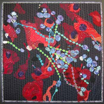
 Branching by Paula Golden Branching by Paula Golden
Nurturing for life begins at the mother’s breast and the importance of breast health cannot be stressed enough. The complex network of tissues that support the formation and delivery of milk in the mammary gland is portrayed via layers of hat and plastic vegetable netting, nylon tulle and commercial fabrics. Machine appliquéd and quilted. |  Branching by Mara Steinkamp, Graduate Student, Human Genetics and Diane Robins, Ph.D., Professor, Department of Human Genetics Branching by Mara Steinkamp, Graduate Student, Human Genetics and Diane Robins, Ph.D., Professor, Department of Human Genetics
The mouse mammary gland consists of milk-producing alveolar cells and a network of ducts that transport the milk to the nipple. Beginning at puberty, ducts grow out from the nipple, invading the surrounding fat pad. Many of the factors important in development of the mammary gland may also be involved in breast cancer initiation and in subsequent tumor growth. In each image, ducts branch into the surrounding fat pads. The colors have been changed in each rendering. |
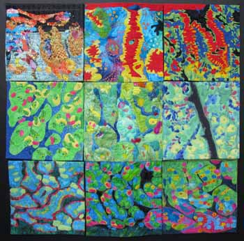 Gastric Rainbow by Carole Nicholas, Judy Busby, Christine Adams, Bunnie Jordan, Paula Golden, Sandi Goldman, Barbara Hollinger, Mary Lois Davis, Annabel Ebersole Gastric Rainbow by Carole Nicholas, Judy Busby, Christine Adams, Bunnie Jordan, Paula Golden, Sandi Goldman, Barbara Hollinger, Mary Lois Davis, Annabel Ebersole
Nine artists independently interpreted a 10″ section. Techniques are varied: fusing, raw edge appliqué, free-motion quilting, machine embroidery, silk waste overlay, zigzagged and turned edges. Materials; 100% cottons (also batiks, and artist dyed), Swarovski beads, acrylic paint, and watercolor pencils. | 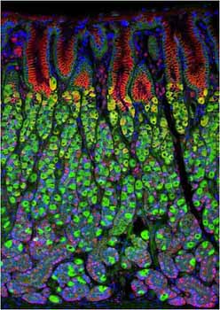 Gastric Rainbow by Jochen Lennerz, M.D., Ph.D., Department of Pathology and Immunology, Washington University School of Medicine, St. Louis, MO and Jason Mills, M.D., Ph.D University of Washington at St. Louis Gastric Rainbow by Jochen Lennerz, M.D., Ph.D., Department of Pathology and Immunology, Washington University School of Medicine, St. Louis, MO and Jason Mills, M.D., Ph.D University of Washington at St. Louis
This is a section of a mouse stomach. Food will be digested in the black area at the top of the picture. The large green cells in the middle of the image produce stomach acid, while the cells at the bottom (highlighted in magenta) make digestive enzymes. The acid and enzymes are squirted up toward the food. The red cells at top line the surface of the stomach and secrete mucus. The mucus barrier protects these beautiful stomach cells from self-digestion. |
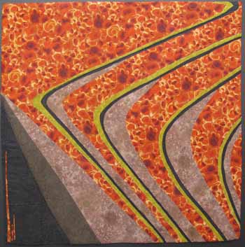 Gradients by Christine Adams Gradients by Christine Adams
The lovely lines of this image remind me of the plains and plateaus in the western part of the USA when viewed from afar. Getting closer we can see all sorts of wild life and flora. Instead of using solids I interpreted this image with a mix of patterned fabrics. It is my way of showing that very little is what it seems from far away. |  Gradients by Edward Flach, Graduate Student, Mathematics Gradients by Edward Flach, Graduate Student, Mathematics
In 1952, Alan Turing proposed that the development of patterns in organisms could be modeled by a mathematical equation. Examples are simple to find: spots on leopards, stripes on fish and shells, even the coloring of pandas! With the possibility that mathematics could help to explain biology, there has been much investigation into this. We have been exploring the model in an abstract way, perturbing Turing’s theory to examine its robustness. Here the horizontal axis is space; the left might be the head of an organism and the right the tail. The vertical axis is time, increasing from bottom to top. The morphogen expression is triggered at the beginning and an oscillatory pattern develops along the entire length of the animal. At high concentration of the morphogen, cells would fix a pigment. |